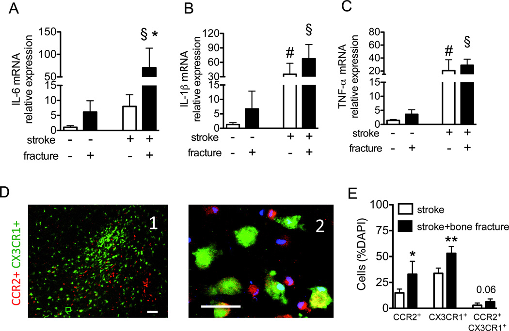
Figure 2. Bone fracture exacerbates brain inflammation.
Relative messenger RNA expression of IL-6 (A), IL-1β (B) and TNF-α (C) six hours after bone fracture and 30 hours after the stroke in the stroke lesion (n=5; (A) *: P=0.01 vs. stroke group without fracture, §: P=0.01 vs. bone fracture group; (B) #: P<0.001 vs. sham procedures for stroke §: P<0.001 vs. bone fracture group; (C) #: P=0.03 vs. the group subjected to sham procedures for stroke and §: P=0.002 vs. bone fracture group). The mice with stroke and bone fracture showed a trend towards higher IL-1β in the brain tissue then the mice with stroke only (P=0.09). D. Representative picture taken in the peri-infarct region of CCR2RFP/+CX3CR1GFP/+ mice with stroke and bone fracture. D1, A low magnified (scale bar: 100 µm) and D2, a high-magnified (scale bar: 50 µm) images show that there are many CCR2+ (red) and CX3CR1+ (green) cells.. Some cells are CCR2-CX3CR1 double positive (yellow). E. The bar graph shows quantification of the percentage of CCR2+, CX3CR1+ or CCR2&CX3CR1+ cells amount total (DAPI positive nuclei) cells in the peri-infarct region (n=5, *: P=0.01 and **: P<0.001). DAPI: 4',6-Diamidino-2-Phenylindole; IL: interleukin; mRNA: messenger ribonucleic acid; TNF: tumor necrosis factor.

