Figure 3.
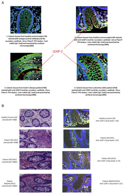
Expression of liver expressed antimicrobial peptide-2 (LEAP-2) protein in the colon tissues. Expression of LEAP-2 protein in the colonic tissue was evaluated using fluorescent immunohistochemistry. (A) Represents the expression of LEAP-2 protein in colonic tissue from normal healthy control subject using anti-LEAP-2 antibody (A2), positive control colon biopsy samples from a patient with Crohn’s disease (A3) or patient with ulcerative colitis (A4) or isotype control (A1) and the secondary antibody Alexa Fluor 594 donkey antirabbit IgG. LEAP-2 appears as red particles in intraepithelial and interstitial tissue of the colon. (B) Shows representatives data of the comparative evaluation of LEAP-2 expression in colonic tissues from a healthy control subject (#105), HIV-monoinfected (#205), HCV-monoinfected (#303) and HIV/HCV-coinfected (#404) patients. The relative expression levels of LEAP-2 for control (#105), HIV patient (#205), HCV-monoinfected patient (#303), and HIV/HCV-coinfected patient (#404) were 7.64, 5.35, 5.14 and 5.61, respectively. The corresponding haematoxylin and eosin (H&E) staining for colonic tissues of the subjects demonstrated an increase in infiltrating inflammatory cells (markers of immune activation) in colonic tissues of patients compared with control.

