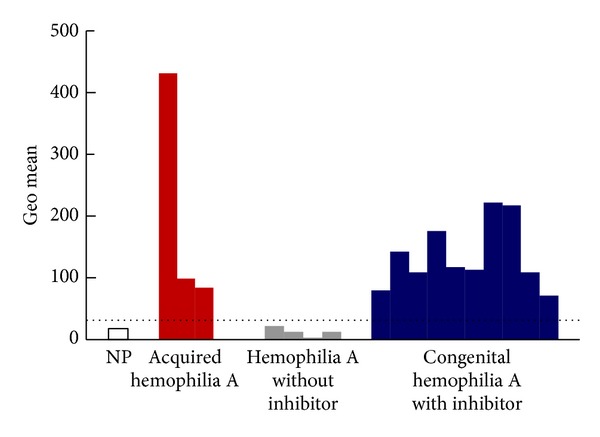
Figure 4.

Flow cytometric analysis of factor VIII- (FVIII-) binding antibodies. Plasma samples from 20 normal healthy volunteers (normal pooled plasma), 3 acquired hemophilia A patients, 4 congenital hemophilia A patients without inhibitors, and 10 congenital hemophilia A patients with inhibitors were assessed using the following procedure. Human recombinant FVIII (rFVIII) was bound to red fluorescent carboxylated polystyrene microbeads (Cyto-Plex polystyrene microbeads) and a certain number of human rFVIII-bound microbeads were added to serially diluted suspected plasma. After incubation and washing, PE-labeled anti-human IgG antibody was added to the microbeads. After additional incubation and washing, fluorescent intensity was measured using a FACScan flow cytometer. The fluorescence intensity of the anti-human IgG antibody bound to human rFVIII on the microbead surface was expressed as the geometric mean (shown in arbitrary units). The dotted line shows a tentative cutoff value for the inhibitor with the highest geometric mean value in plasma without inhibitor. NP: normal plasma.
