
Figure 3.
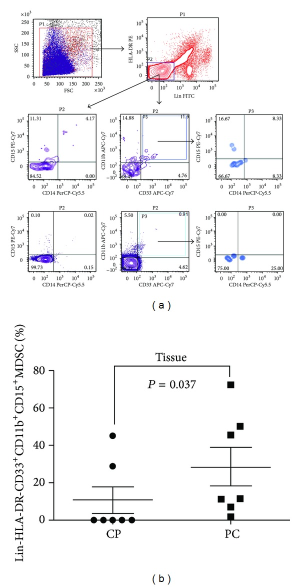
Levels of tumour-infiltrating MDSCs in patients with pancreatic cancer compared to chronic pancreatitis patients. (a) Flow cytometric evaluation of Lin, CD33, CD11b, CD15, and CD14 in tissue samples. An example of representative dot plots is shown for each study sub-group. Gates were set based on controls. Numbers represent the percentages from the original populations gated. P (number) above each FACS plot indicates the population gated that was analysed. The axis of each FACS plot represents the marker analysed. (b) Scatter plot of the percentage of Lin-HLA-DR-CD33+CD11b+CD15+ in the tissue of benign and cancer samples. Bar represents median in each group. CP: chronic pancreatitis; PC: pancreatic cancer.