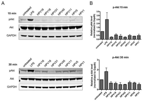
Fig. 4. Disruption in the Akt signaling pathway in LC is similar after treatment with high- and low-risk mucosal HPV genotypes and cutaneous HPV genotypes.
(A) Cellular extracts from LC exposed to HPV genotypes for 15 min (top) or 30 min (bottom) at 37°C were separated by SDS-PAGE, transferred to PVDF and immunoblotted using antibodies to p-Akt (Ser473), total Akt, and GAPDH. Visualization was performed using the Odyssey Infrared Imaging System. (B) Protein bands were quantified using the Odyssey system software, normalized to total Akt and GAPDH expression, and the relative levels of pAkt (mean ± SEM) compared to untreated LC are shown from three individual experiments.

