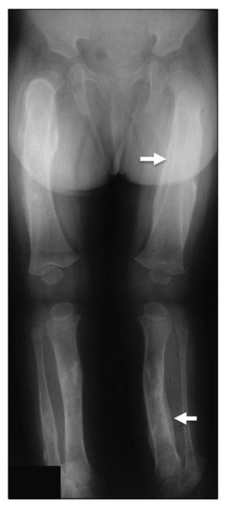
Figure 1. Radiographic features of Caffey disease.

Representative radiograph of an affected individual showing periosteal thickening of the femur and the tibia (white arrow). Adapted from Gensure et al., 2005 [20].

Representative radiograph of an affected individual showing periosteal thickening of the femur and the tibia (white arrow). Adapted from Gensure et al., 2005 [20].