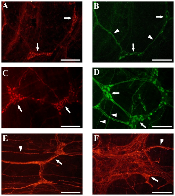
Figure 6. Representative images of colon whole mounts in En/En and en/en rabbit myenteric plexuses.
A) En/En myenteric ganglia (arrows) with human neuronal protein immunoreactive (HuC/D-IR) neurons. B) En/En myenteric ganglia (arrows) with nitric oxide synthase immunoreactive (nNOS-IR) neurons, (arrowheads) nNOS-IR nerve bundles. C) en/en myenteric ganglia (arrows) with HuC/D-IR neurons. D) en/en myenteric ganglia (arrows) with nNOS-IR neurons, (arrowheads) nNOS-IR nerve bundles arranged within primary, secondary and tertiary nerve strands. In En/En (A and B) the ganglia and nerve bundles are less dense, have a lower number of HuC/D-IR and nNOS-IR neurons compared to en/en rabbits (C and D). E) and F): differences between En/En vs en/en SP-IR neurons (arrow), ganglia and nerve bundles (arrowhead) in the morphology of myenteric plexus in the ascending colon. In En/En (E) ganglia (arrow) are small and have a lower number of substance P immunoreactive (SP-IR) neurons than en/en rabbits (F). A–D scale bars 200 µm; E–F scale bars 100 µm.

