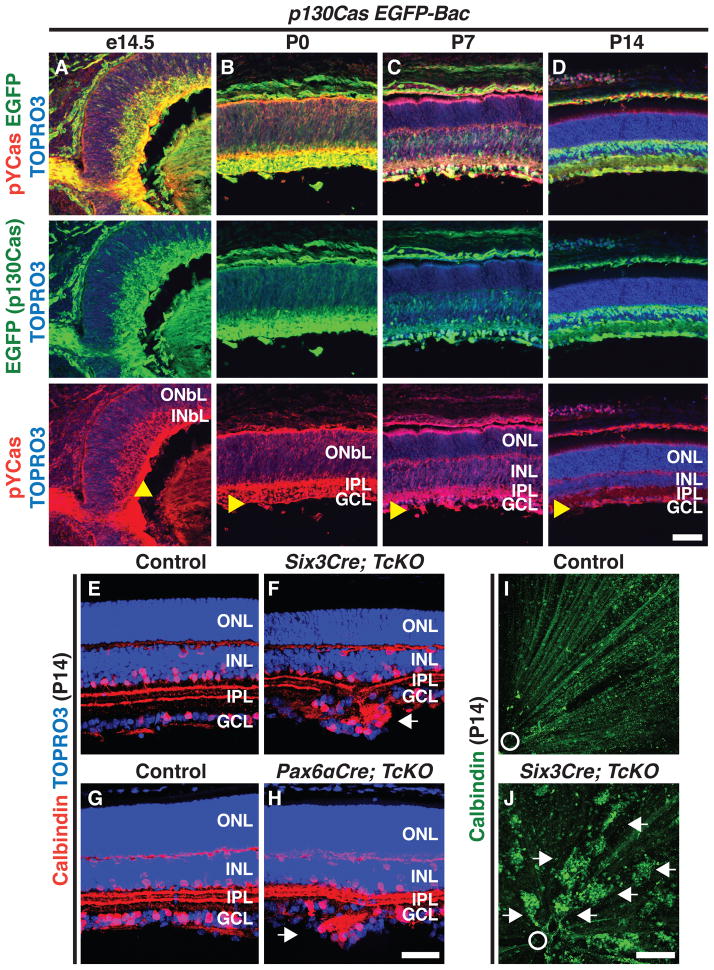
Figure 2. Cas Signaling Adaptor Proteins are Required for the GCL to Resolve into a Single Cell Layer.
(A–D) Expression of a p130Cas EGFP reporter (EGFP, left green) and phosophorylated-p130Cas (PY-Cas, red) in a p130Cas EGFP Bac transgenic mouse (p130Cas EGFP-Bac) throughout retina development. Retinas were counterstained with TOPRO3 (blue). From e14.5 to P0 (A and B) p130Cas phosphorylation is mainly found in the INbL and is enriched in close proximity to the ILM. (E–H) Cryo-sectioned Control (E, G), Six3Cre; TcKO (F) and Pax6αCre; TcKO (H) retinas immunostained with anti-calbindin (red) and TOPRO3 (blue). Retina-specific removal of Cas (F and H) results in the formation of ectopic cell aggregates in the GCL beyond the ILM (n=5 independent animals for each genotype). (I and J) Whole-mount calbindin immunostaining (green) confirms the presence of ectopic GCL cell aggregates throughout the retina in Six3Cre; TcKO (J) mice, as compared to Control (I). Arrowheads: ILM; white arrows: ectopic aggregates; white circle: optic nerve head. Scale bars, 50 μm for A–D and E–H, and 200 μm for I and J.

