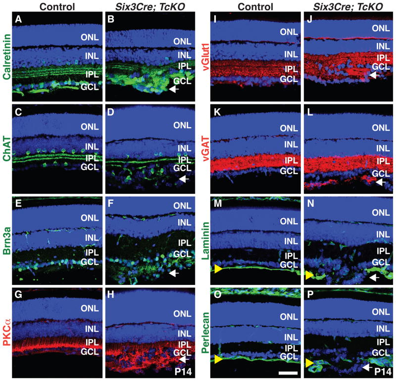
Figure 3. Ectopic GCL Aggregates in Cas TcKO Mutant Retinas are Formed by Multiple Cell-types.
(A–P) Control (A, C, E, G, I, K, M and O) and Six3Cre; TcKO (B, D, F, H, J, L, N and P) P14 retina sections were immunostained with antibodies against the AC and RGC marker calretinin (A and B), the starburst AC marker ChAT (C and D), the RGC marker Brn3a (E and F), the rod BP cell marker PKCα (G and H), the presynaptic terminal markers vGlut1 (I and J) and vGAT (K and L), and the ILM markers laminin (M and N) and perlecan (O and P). Six3Cre; TcKO ectopic aggregates contain both RGCs and displaced ACs (B, D and F). Note that the ILM is completely disrupted at sites where aggregates form in the Six3Cre; TcKO (N and P) retinas compared to control (M and O). All sections were counterstained with TOPRO3 (Blue). White arrows: ectopic aggregates; yellow arrowheads: ILM. Scale bar, 50 μm.

