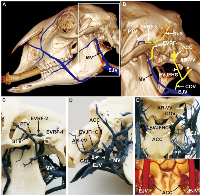
Figure 4. Course of the emissary veins of the temporal sinus and the formation of the anterior condylar confluent.
(A) 3D CT reconstruction of the head combined with schematic vein drawings (blue), lateral, left view. White frame: inset of B. The ovine extracranial veins can be observed in this view, particularly the outer drainage system of the intracranial veins. (B) 3D CT scan combined with schematic diagram of blue and orange colored veins (interrupted orange vein of temporal sinus shows the invisible part of the sinus in the temporal meatus), lateral left view (paracondylar process removed). (C) Corrosion cast, lateral left view. The temporal sinus (TeS) ran through the temporal meatus and split into two distinct vessels, (1) the first emissary vein (EVRF-1), which left the main opening of the retroarticular foramen and joined the maxillary vein (MV), and (2) the second emissary vein (EVRF-2) which ran next to a tributary canal, passed a tributary foramen, and joined the profundal temporal vein (PTV). (D) Corrosion cast, lateral right view. (E) Corrosion cast, ventrolateral view. The emissary vein of the jugular foramen and the emissary vein of the hypoglossal canal converged towards an extracranial orifice and formed the ‘anterior condylar confluent’ (ACC). The emissary vein of the jugular foramen and hypoglossal canal (EVJFHC) merged with an anastomotic ramus of the vertebral vein (AR-VV), to form the craniooccipital vein (COV), which drained into the external jugular vein (EJV). (F) 3D CT scan, ventrolateral view. The ACC is a clearly visible structure in the sheep. ACC: anterior condylar confluent, AR-VV: anastomotic ramus of the vertebral vein, COV: craniooccipital vein, EJV: external jugular vein, EVJFHC: emissary vein of the jugular foramen and hypoglossal canal, EVRF-1: first emissary vein of retroarticular foramen, EVRF-2: second emissary vein of retroarticular foramen, MV: maxillary vein, PP: pterygoid plexus, PTV: profundal temporal vein, STV: superficial temporal vein, TeS: temporal sinus.

