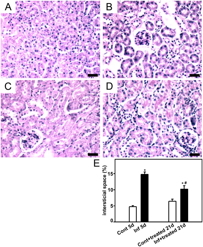
Figure 5. Effect of antimalarial treatment on the size of the interstitial space.
Animals were euthanized on day 5 or 21 post infection (p.i.), perfused, and the kidneys were collected for histological analysis. The size of the interstitial space in the renal cortex was visualized using periodic acid−Schiff. Representative photomicrography of noninfected mice (control5d) (A), 5 days p.i. (infected5d) (B), noninfected mice that received treatment with antimalarial drugs (control+treated21d) (C), and infected mice that received treatment with antimalarial drugs (infected+treated21d) (D) (n = 6 per group). Bar = 20 µm. The size of the interstitial space is quantified in (E). Values are expressed as a percentage of interstitial space per area (means ± standard error). *Statistically significant compared with control5d or control+treated21d. #Statistically significant compared with control5d or infected5d (P<0.05).

