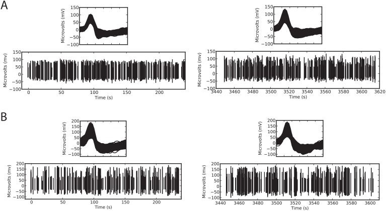
Figure 2. Comparison of extracellular recording using PriED and a similarly designed professionally machined microdrive.
Recordings were made using 700–900 k FHC tungsten electrodes. (A) Sample recording using the stacked base design and using a cannula to pass the electrode through the dura to the anterior cingulate cortex of an awake NHP. (B) Sample recording using a professionally machined version of the microdrive recording from the basal forebrain in an awake NHP. Both panels show two excerpts from the recordings: 500 action potentials from the start of recording (near time 0s) and 500 action potentials from the end of the session (around 3500s). The insets show details of the recorded action potentials.

