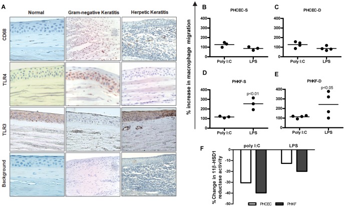
Figure 4. Macrophage infiltration in human keratitis.
(A) Immunohistochemistry of the normal human central corneal epithelium showed no evidence of CD68 positive resident macrophages or basal TLR4 expression, although there was some basal TLR3 expression. Corneal stromal infiltration of CD68 positive cells is seen in both herpetic and gram negative keratitis associated with increased TLR3 and TLR4 expression in the corneal epithelium, respectively. (B–E) Migration assay showing culture supernatants of corneal cells having chemotactic potential on monocytes. Cell supernatants from PHCEC/PHKF stimulated with TLR3 (poly I∶C) or TLR4 (LPS) ligands for 16 h were tested for the ability to induce monocyte migration. ‘S’ denotes culture supernatants from PHCEC/PHKF cultures generated from 3 corneal donors, tested on a single allogenic PBMC donor. ‘D’ denotes 3 different PBMC donors subjected to culture supernatant from a single donor derived PHCEC/PHKF treated with TLR3 and TLR4 ligands. Data show that both LPS and Poly I∶C stimulation of corneal cells induce monocyte migration but LPS stimulation of PHKF has the greatest chemotactic potential. (Panels D and E). (F) Culture supernatants from experiments A–D (TLR3 and TLR4 induction of PHCEC/PHKF for 16 h) downregulates M1 macrophage 11β-HSD1 activity. Statistical analysis was carried using one-way ANOVA and comparisons were drawn with untreated control cells vs. TLR3/TLR4 treated cells.

