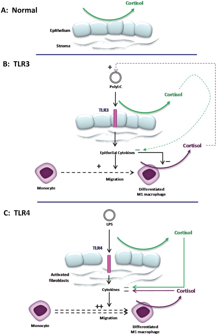
Figure 7. Putative interaction of TLR signaling and local regulation of cortisol in the human cornea.
(A) Under physiological conditions, autocrine synthesis of cortisol in corneal epithelial cells contributes to the immunoprotection of the ocular surface mucosa. During induction of ocular surface TLRs cytokines are released in a ligand and cell specific manner. (B) On TLR3 ligation such as chronic immune -mediated disease e.g. SJS-TEN, synthesis of a diverse spectrum of cytokines primarily from the corneal epithelial cells, induces weak monocyte chemotaxis and differentiation to M1 macrophages. These cytokines potently attenuate M1 cortisol biosynthesis leading to a net reduction of ocular surface cortisol levels, promoting recruitment of inflammatory cells necessary for resolving the initial trigger. By contrast, on TLR4 ligation (C) activation of keratocytes to a fibroblast phenotype, form the first line of defense producing chemokines that potently induce monocyte migration to the site of infection for rapid eradication of bacterial invasion. Attenuation of M1 cortisol production is less pronounced and this facilitates resolution of the inflammatory response, limiting tissue damage thereby preserving optical clarity (and sight).

