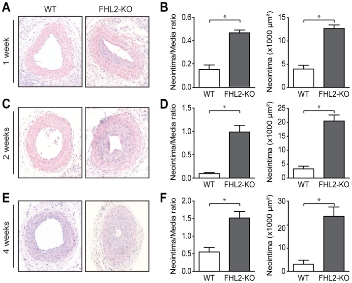
Figure 1. Deficiency of FHL2 accelerates neointima formation after carotid artery ligation.
A, C and E; Representative cross sections of hematoxylin/eosin-stained carotid arteries from WT and FHL2-KO mice ligated for 1 (A), 2 (C) and 4 weeks (E). B, D and F; Quantitative analysis of neointima/media ratio and neointimal area in histological sections from WT and FHL2-KO mice ligated for 1 (B), 2 (D) and 4 weeks (F), revealed increased lesion formation in FHL2-KO mice. n = 7 for 1 and 2 weeks and n = 14 for 4 weeks. Three consecutive sections per mouse at each location were employed in the analysis. Lesions were characterized at 1.7, 2.0 and 2.3 mm from the reference point at 1, 2 and 4 weeks, respectively. Values are mean±SEM. *P<0.05 for FHL2-KO versus WT mice.

