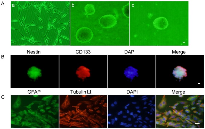
Figure 1. Isolation and identification of GSCs.
(A) a: U87 cell lines attached and grew as a monolayer in flasks. b: The spheres formed and reached 100–200 cells each in the serum-free medium. c: Subsphere-forming assay of single-cell suspensions from the cell spheres. (B) Immunofluorescence staining of Nestin (green) and CD133 (red) in cell spheres. (C) Glioblastoma sphere-differentiated progenies were stained with GFAP (green) and beta-tubulin III (red). Scale bars represent 20 µm.

