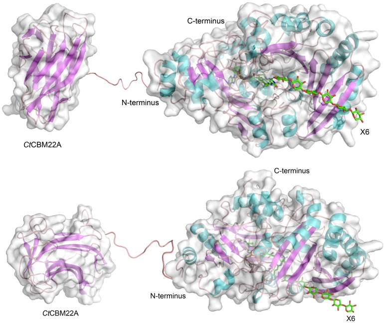
Figure 1. Structural model of the fusion protein.
GOOX-VN was fused to C. thermocellum CBM22A (PDB ID: 1DYO) via a TP-rich linker. The model of GOOX-VN was built from the X-ray structure of GOOX-T1 (PDB ID: 2AXR) while the non-helical model of the linker was built from the X-ray structure of a Bacillus subtilis polysaccharide deacetylase (PDB ID: 1NY1). Xylohexaose (X6) was docked into the active site of GOOX-VN using Autodock 4. Top: the substrate-entrance view of the fused protein model; Bottom: the side view of the fused protein model.

