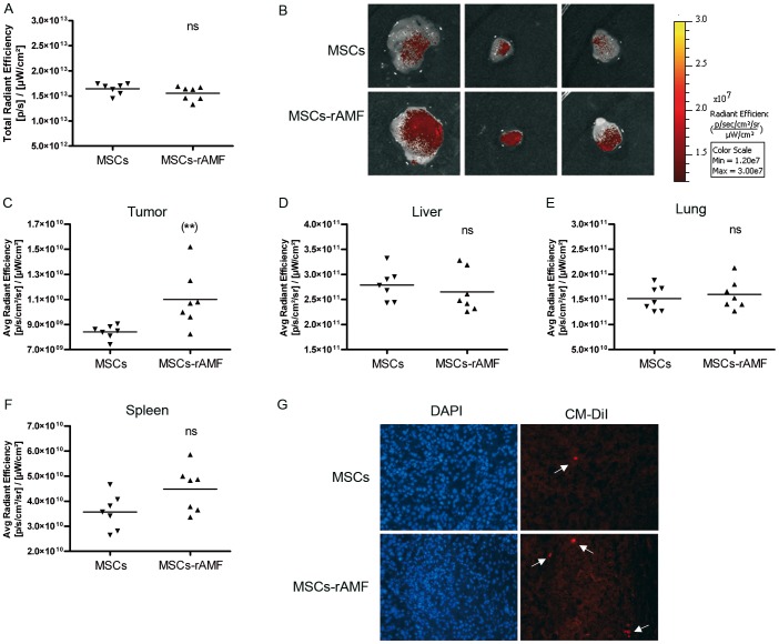
Figure 5. rAMF increases the in vivo migration and anchorage of MSCs to HCC tumors.
BM-MSCs prestimulated with 1 µg/ml of rAMF were labeled with DiR and CMDiI cell trackers and IV injected in SC HuH7 tumor-bearing mice. After 3 days, tumors were removed and exposed to FI. A) Total FI was calculated by measuring the region of interest (ROI) for all the tissues isolated and the results were expressed as total radiant efficiency. ns, non significant. B) Representative tumor images of mice inoculated with rAMF-prestimulated BM-MSCs (MSC-rAMF) or unstimulated cells (MSCs). Images represent the average radiant efficiency. Region of interest (ROI) was calculated for the isolated tumor (C), liver (D), lung (E) and spleen (F) and the results were expressed as the average radiant efficiency. **p<0.01 vs unstimulated BM-MSCs (unpaired Student's t-test). G) Microscopic analysis of transplanted CM-DiI-labeled MSCs (red signal indicated by arrows) and DAPI staining in frozen sections of tumors. Magnification ×200.

