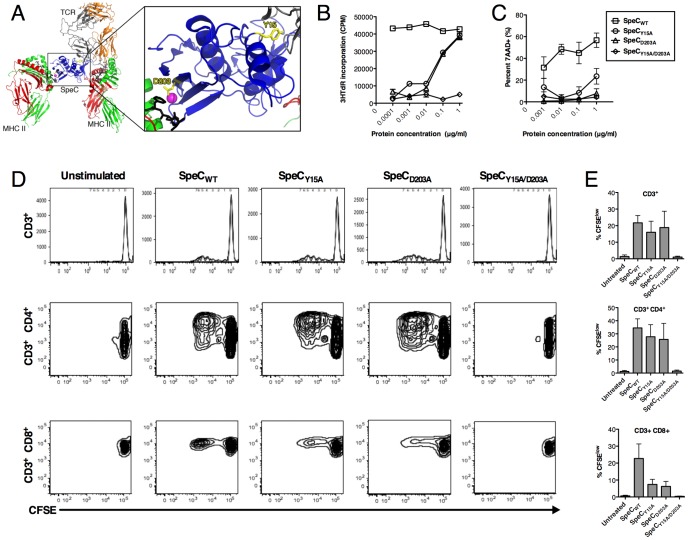
Figure 1. Overview of the SpeC-mediated T cell activation complex and mutations to reduce systemic toxicity.
A) Structural overview of SpeC in complex with TCR and MHC-II. TCR Vα chain is colored orange, TCR Vβ chain is colored grey, MHCα-chains are colored red, MHCβ-chains are colored green, antigenic peptides are colored black, and the zinc atom is colored magenta. SpeC is colored blue with important interface residues Y15 and D203 highlighted in yellow. The ternary model of TCR-SpeC-(MHC)2 was produced as described previously [9] and the ribbon diagram was generated using PyMOL (http://www.pymol.org). B) Proliferation of human PBMCs mediated by SpeCWT or proteins containing mutated residues Y15A (TCR-binding mutant), D203A (MHC-II-binding mutant) or Y15A/D203A was determined by the uptake of 3H-thymidine after 72 h post-stimulation (n = 5 in triplicate; data representative of one individual). C) Dose-dependent cytotoxicity of 7-AAD+ WiDr cells after 48 h incubation with human PBMCs and either SpeCWT, SpeCY15A, SpeCD203A, or SpeCY15A/D203A (n = 3–6 per group). (D–E) Proliferation of CFSE labeled-human PBMCs mediated by SpeCWT or proteins containing mutated residues was determined by FACS five days post-stimulation, specifically measuring total CD3+ T cell population, CD3+CD4+ T cells and, CD3+CD8+ T cells (n = 4; FACS data representative of one individual).

