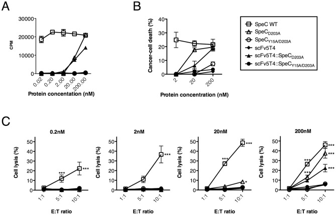
Figure 4. Functionality of SpeC mutants and fusion proteins for human PBMC proliferation and cytotoxicity in vitro.
A) SpeC proteins were used to compare scFv5T4 alone, scFv5T4::SpeCY15A/D203A and subsequently scFv5T4::SpeCD203A in the uptake of 3H-thymidine as a measure of PBMC proliferation after 4 day incubation (n = 5). B–C) Dose-dependent SpeC-mediated PBMC cytotoxicity of scFv5T4::SpeCD203A was determined by comparing SpeC controls, scFv5T4 alone and scFv5T4::SpeCY15A/D203A after 48 h incubation by using FACS analysis of WiDr (panel B), measuring percent cancer cell death with 7AAD-exclusion staining (n = 3) and 51Cr-release to measure the specific cytotoxic potential (panel C) when incubated with increasing effector∶target ratios and 51Cr-labeled HT-29 cancer cells. Data shown (mean ±SEM) is from four independent human donors each done in triplicate. *p<0.05, ***p<0.001, compared to the inactive SpeCY15A/D203A control protein.

