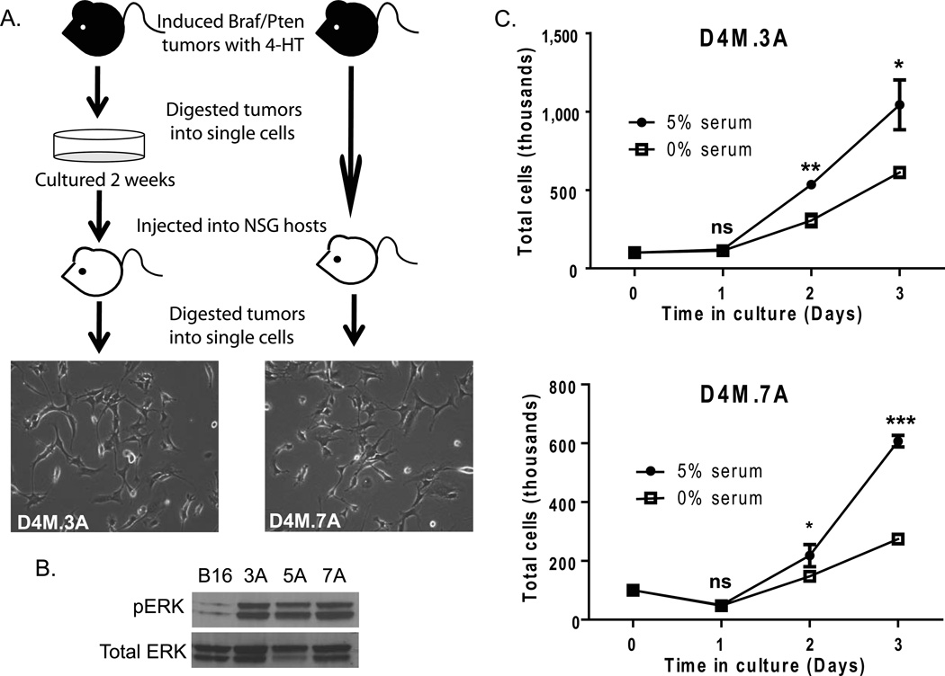
Figure 1. D4M cell lines grow readily in culture and exhibit constitutive activation of pERK.
(A) Schematic of the protocol used to generate D4M cell lines. Braf/Pten mice were injected with 4-hydroxytamoxifen (4-HT) and the tumors that developed were excised and dissociated into single cells. These cells were cultured for 2 weeks (left) or uncultured and immediately injected intradermally into NSG host mice after dissociation (right). Tumors that arose from the host mice were excised and dissociated into single cells and plated in vitro to generate D4M cell lines. Representative brightfield images of D4M.3A and D4M.7A were taken at 10× magnification. (B) Immunoblot analysis for pERK of 4µg of protein lysate from B16, D4M.3A, D4M.5A, and D4M.7A cultured cells. Total ERK was used as a loading control. (C) In vitro growth curves of D4M.3A and D4M.7A cells in DMEM/F-12 advanced with either 0% or 5% FBS. Cells, 100,000, were plated in triplicate wells and total numbers of cells were counted over the next 3 days. Error bars represent standard deviations of the means. T-tests were performed on the averages of 4 separate wells for each time point (ns = not significant, * = P < 0.05, ** = P < 0.005).

