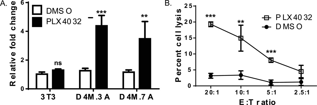
Figure 5. PLX4032 increases functional pmel expression in D4M cells.
(A) RT-PCR comparing pmel expression in mouse 3T3, D4M.3A, and D4M.7A cell lines. Cells were plated in triplicate wells and treated with either DMSO or 3µM PLX4032 for 48 hrs. 3T3 cells were used as a negative control. Samples were normalized to Gapdh and fold change was calculated relative to 3T3 cells treated with DMSO. T-tests were performed on the averages of the triplicate wells, relative to DMSO treatment in each cell line. Error bars represent standard deviations of the means. (B) Chromium release assay of D4M.3A cells treated with either DMSO or 3µM PLX4032 for 72 hrs. Pmel transgenic T cells were incubated with D4M.3A cells for 4 hours and cell lysis was plotted with the different Effector to Target (E:T) ratios. Error bars represent standard deviations. T-test results are represented as: ns = not significant, * = P < 0.05, ** = P < 0.005, *** = P < 0.0005).

