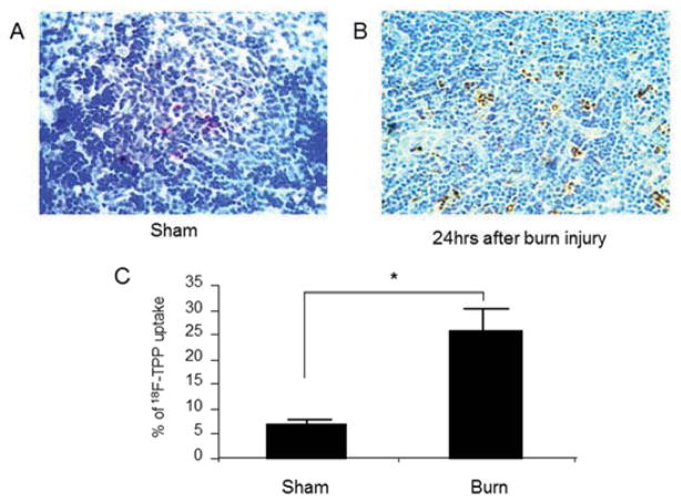
Fig. 4. Burn induced apoptosis in mouse spleen.
TUNEL staining on the sections of the spleens from the sham mice demonstrated diffusely scattered apoptotic cells at a rate of 4.4 +/−1.8% in the white medulla (Fig. 4 A). By contrast, burn induced significant cellular apoptosis in the spleen presented an apoptotic index of as high as 24.6+/− 6.7 % (Fig. 4 B, C: sham vs. burn: t test, p<0.005). The majority of the apoptotic cells were present in the white medulla area.

