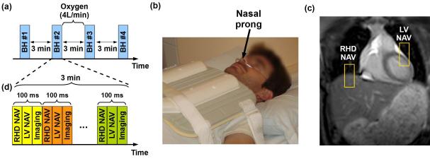
Figure 1.
Imaging protocol for breath-hold (BH) assessment. (a) Study protocol designed for BH assessment without (BH#1, BH#2, and BH#4) and with (BH#3) supplemental oxygen and hyperventilation. Oxygen (4 l/min) was administrated using nasal prolong as illustrated in (b), (c) and (d) show the sequence diagram used for BH monitoring. A real time steady-state free precession (SSFP) sequence was used to acquire a complete image every 100ms using a right hemidiaphragm (RHD) navigator (NAV), a NAV located at the left ventricle LV (LV NAV) and a 2D coronal imaging slice (Imaging) (d).

