Figure 2.

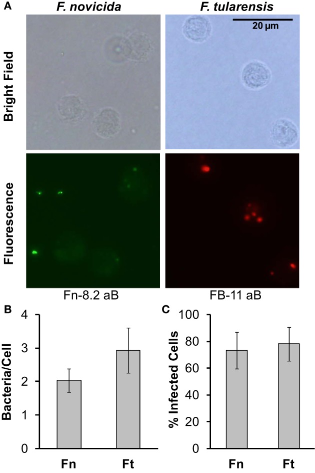
F. novicida and F. tularensis associate similarly with monocytes during infection. Primary human monocytes were infected with F. novicida (Fn) or F. tularensis Schu S4 (Ft) at an MOI of 50 for 5 h. (A) Representative images from cells fixed in paraformaldehyde and then stained with Fn-82 antibody specific for F. novicida or FB-11 antibody specific for F. tularensis. The corresponding secondary antibody for Fn-82 was Alexa Fluor 488 rabbit anti-goat IgG (green) and for FB-11 it was AlexaFluor 594 goat anti-mouse IgG (red). Graphs represent the number of bacteria per cell (B) or the number of infected cells (C). Graphs represent the mean ± s.e.m. from 1 donor incorporating a minimum of 4 frames. Data were analyzed by Student's t-test. No significant differences were found.
