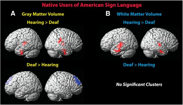
Figure 1.
GMV and WMV differences between deaf and hearing native users of ASL. Less GMV (A) was observed in the deaf group in bilateral temporal lobe regions, including Heschl's gyrus. Less WMV (B) was also observed in the bilateral temporal lobe regions, close to areas identified to differ in gray matter. Deaf signers had more GMV in right superior frontal cortex. Height threshold p < 0.005; nonstationary corrected threshold p < 0.05. Clusters were overlaid onto the standardized MNI brain template.

