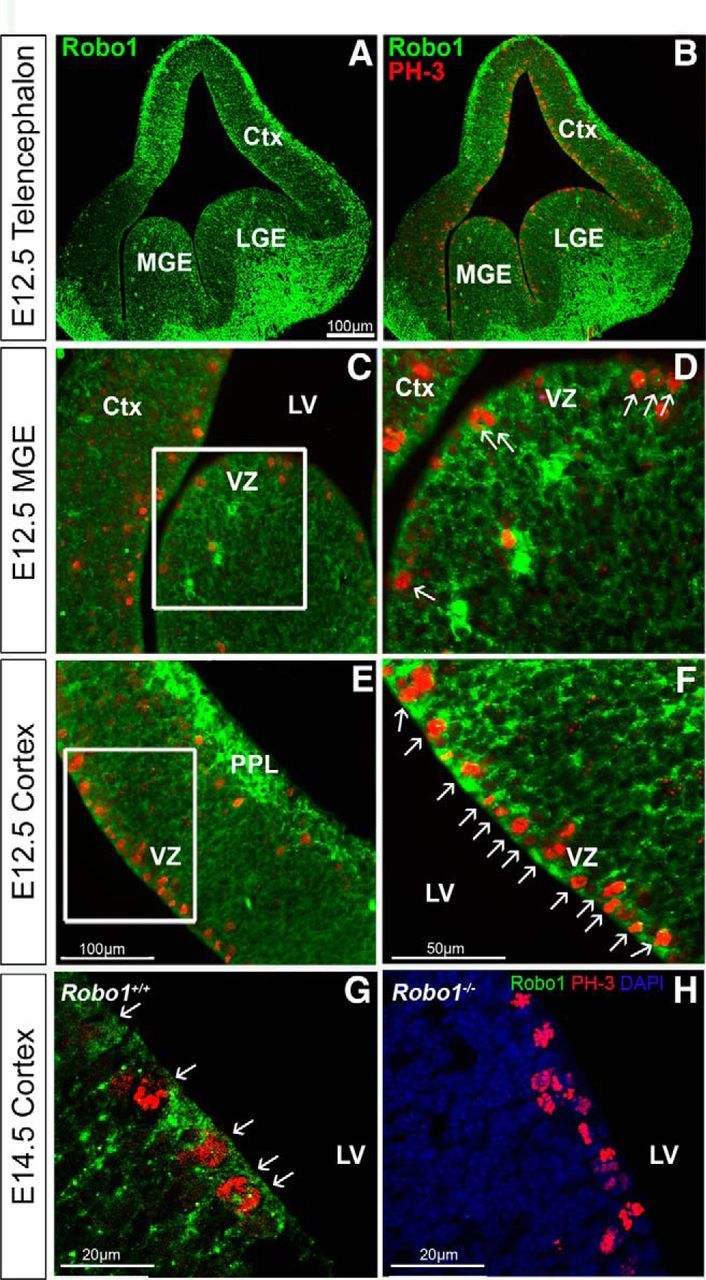
Figure 2.

Coexpression of Robo1 protein with the proliferation marker, PH-3. Immunohistochemical localization of Robo1 (green) and PH-3 (red) in coronal sections through the telencephalon (A, B), and through the VZ of the MGE (C, D) and cortex of C57BL/6J mice at E12.5 (E, F), and in Robo1+/+ (G) and Robo1−/− littermate (H) at E14.5. D, F, Higher-magnification of the boxed areas in C and E, respectively, and illustrate coexpression of the two markers (arrows) in individual cells of the VZ of the MGE (D) and in the majority of the cells in the VZ of the cortex at both ages (F, G). Ctx, Cortex; LV, lateral ventricle.
