Figure 5.

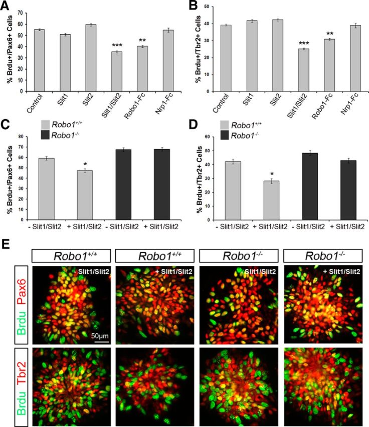
Reduced proliferation in Robo1+/+, but not in Robo1−/− dissociated cortical cell cultures following Slit1/Slit2 treatment. A, B, Histograms show percentage of BrdU+ E15.5 rat apical (Pax6+; A) and basal (Tbr2+; B) progenitor cells following treatment with either: Control, Slit1, Slit2, Slit1/Slit2, Robo1-Fc, or Nrp1-Fc overnight. Treatment with Slit1/Slit2 and Robo1-Fc caused a significant decrease in proliferation of both progenitor pools. C, D, Dissociated cortical cell cultures prepared from E13.5 Robo1+/+ and Robo1−/− mice were incubated overnight in the presence or absence of Slit1/Slit2, and the percentages of proliferating apical (C) and basal (D) progenitors were assessed following a 2 h BrdU pulse. Wild-type cultures showed a significant decrease in proliferation following Slit1/Slit2 treatment. Robo1−/− dissociated cortical cell cultures were unaffected by Slit treatment, but showed increased proliferation compared with wild-type cultures (E); *p < 0.05, **p < 0.004, ***p < 0.0004.
