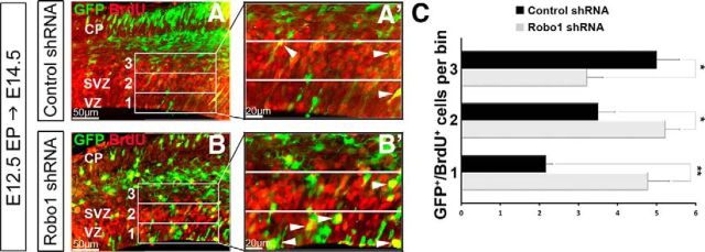
Figure 9.
Increase in proliferative activity in the absence of Robo1 is cell autonomous. (A–B′) Immunohistochemistry in the dorsal cortex of C57BL/6J mice at E14.5, 48 h after in utero electroporation of either control shRNA (A, A′) or Robo1shRNA (B, B′) for GFP+ (green, electroporated) and BrdU+ (red) cells, following a 2 h BrdU pulse before kill. C, Histogram shows a significant increase in proliferating Robo1 shRNA electroporated cells (GFP+/BrdU+) present in lower bins within the VZ and SVZ of the cortex compared with control shRNA electroporated embryos (A′, B′); *p < 0.05, **p < 0.004.

