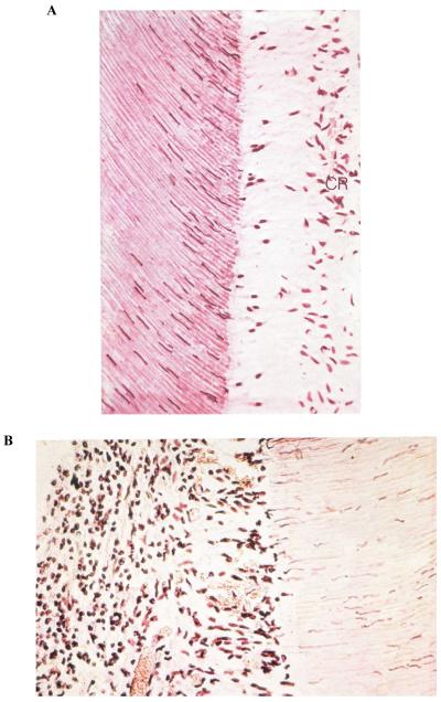
Fig. 4. Histologic appearance of young dental pulp.
(A) After evaporative air blast to exposed dentine surface in vivo. Note the loss of the odontoblastic cell bodies and the appearance of their nuclei in the adjacent dentinal tubules. The cell-free zone is seen just above the cell-rich (CR) zone. There are no inflammatory cells in this pulp. (B) After exposed coronal dentine was left open to oral fluids for 1 week. The pulp is heavily inflamed. This dentine was very hypersensitive. Note the absence of an odontoblast layer and the cell-free and CR zones, and the presence of acute inflammatory cells throughout the pulp. A large dilated venule can be seen in the bottom of the field.
Reproduced from Brännström,1 with kind permission from Elsevier.

