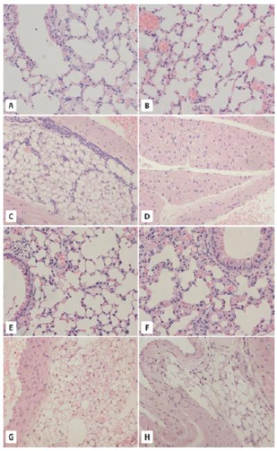
Fig. 1.

The morphology of lung- and aorta-associated adipose tissue in GHR-KO and bGHTg mice. A and B. Morphology of the lungs of WT (A) and GHR-KO (Laron dwarf) mice (B). The pulmonary alveoli of 2-year-old WT mice and 2-year-old GHR-KO mice show a normal structure of terminal bronchioles and alveoli. C and D. Analysis of adipose tissue from the aorta region of WT (C) and Laron dwarf mice (D). Adipose tissue sections from 2-year-old WT mice (C) reveal the presence of white adipose tissue (WAT) only and leukocyte infiltration. By contrast, 2-year-old GHR-KO mice (D) show the presence of brown adipose tissue (BAT) with no leukocyte infiltration. E and F. Lung morphology in 6-month-old WT (C) and bGHTg (F) mice. Both alveoli and terminal bronchioles show normal structure. G and H. Analysis of adipose tissue from the aorta region of WT (G) and bGHTg (H) mice. While the 6-month-old WT mice have both WAT and BAT tissue visible around the aorta (G), the 6-month-old bGHTg mice have WAT with only a few brown adipocytes visible. × 200.
