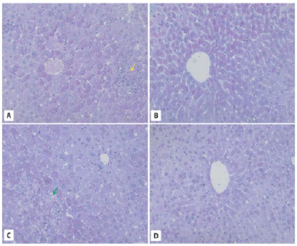
Fig. 2.

Liver morphology in 2-year-old WT and GHR-KO mice. A, C. PAS staining of liver sections from WT mice reveals abundant lipid droplets forming macrovesicles (red arrows), fat microvesicles containing ballooning cells, leukocyte infiltrations (yellow arrow), and the presence of hepatocytes with intranuclear inclusions (green arrow). B, D. Liver sections in 2-year-old GHR-KO mice show normal morphology. × 200.
