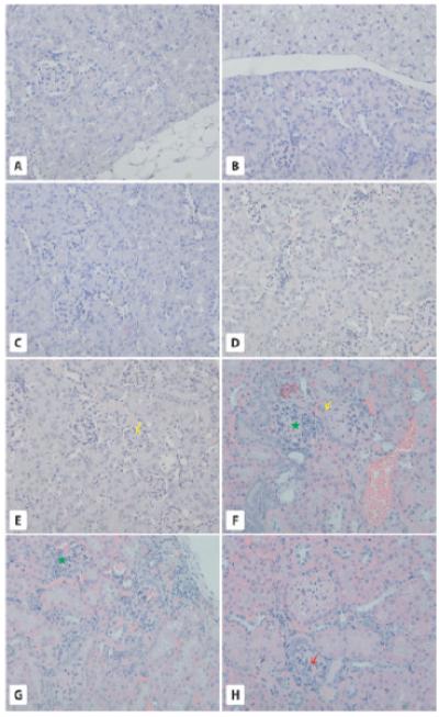
Fig. 4.

Morphology of the kidneys. Panel A-D. Two-year-old WT and GHR-KO mice. While kidneys in 2-year-old WT mice are surrounded by WAT (A) kidneys in GHR-KO mice are surrounded by both WAT and BAT (B). Overall, kidneys in WT (C) and GHR-KO (D) mice show normal morphology. E-H. Six-month-old WT and bGHTg mice. While 6-month-old WT mice show normal structure for the renal cortex (E), the yellow arrow points to a normal glomerulus), 6-month-old bGHTg mice show several structural changes in kidneys, such as a transformation from simple to stratified epithelium on the outer layer of Bowman’s capsule (F and H, yellow arrows), leukocyte infiltration (F, green asterisk), damaged glomeruli with leukocyte infiltration (G, green asterisk) and signs of glomeruosclerosis (H, red arrow). × 200.
