
Figure 3. Binding of CPMV conjugates to endothelial cellsin vitro.
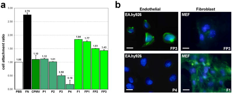
a. Cell adhesion assay with human endothelial cells (AE.hy926). Results are normalized to PBS control (y axis, Binding Efficiency: relative binding, normalized to PBS). Assay was performed in duplicate. Positive control: fibronectin (FN); negative control: native CPMV (CPMV); see Table 1 for an explanation of CPMV conjugates. b. Fluorescence visualization of CPMV conjugates FP3 (loaded with PEG500f and F56f), P4 (loaded with PEG500f only) and F1 (loaded with F55f only) bound to endothelial cells (EA.hy926) and fibroblasts (MEF). CPMV particles are shown in green. Nuclei were stained with DAPI (in blue).