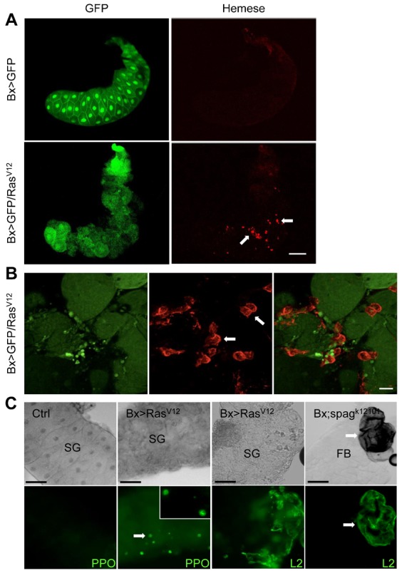
Fig. 3. RasV12-expressing salivary glands are infiltrated by hemocytes.

(A) Overview of glands from RasV12 and control larvae. GFP-expressing control glands and RasV12-expressing glands (the left part shows the GFP signal) labeled with a hemocyte-specific antibody (anti-Hemese, right panel, some hemocytes are indicated by arrows). (B) Hemocytes (arrows) spread around RasV12 gland cells. A detailed view of a RasV12-expressing gland such as in panel A is shown. (C) Crystal cells and lamellocytes attach to RasV12-expressing glands. Left part: glands (SG) from a control cross (Bx-GAL4;+/+) and RasV12-expressing glands were labeled with a prophenoloxidase-specific antibody and visualized using epifluorescence. Right part: lamellocytes (L2) were visualized using a specific antibody (Kurucz et al., 2007) in RasV12-expressing glands (left) and the fat body (FB) of an autoimmune mutant, which was used as a positive control (spaghetti, spagk12101). Scale bars: 100 µm (A), 20 µm (B), 50 µm (C).
