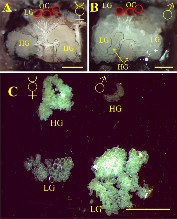
Fig. 3. Hypopharyngeal glands in bumblebee males.

Frontal cuticle was removed from the heads of B. terrestris worker (A) and drone (B). The underlying space was mainly filled with labial glands (LG) with large acini in males and with hypopharyngeal glands (HG) in females. In the central part of the male head the distinct glandular tissue of HGs with brownish duct and small acini could be observed (dashed lines). Hypopharyngeal glands were uncovered from the overlaying LG tissue by forceps. Red circles in both pictures indicate the positions of ocelli (OC). (C) Dissected head glands of a worker (left) and a drone (right). Labial glands (LGs) fill the posterior space of the head in both males and females. Anterior part of the male's head is filled by large LGs hiding small HGs. Scale bars: 1 mm (A,B), 2 mm (C).
