
Fig. 5. F-actin rings of bumblebee and honeybee secretory cells.
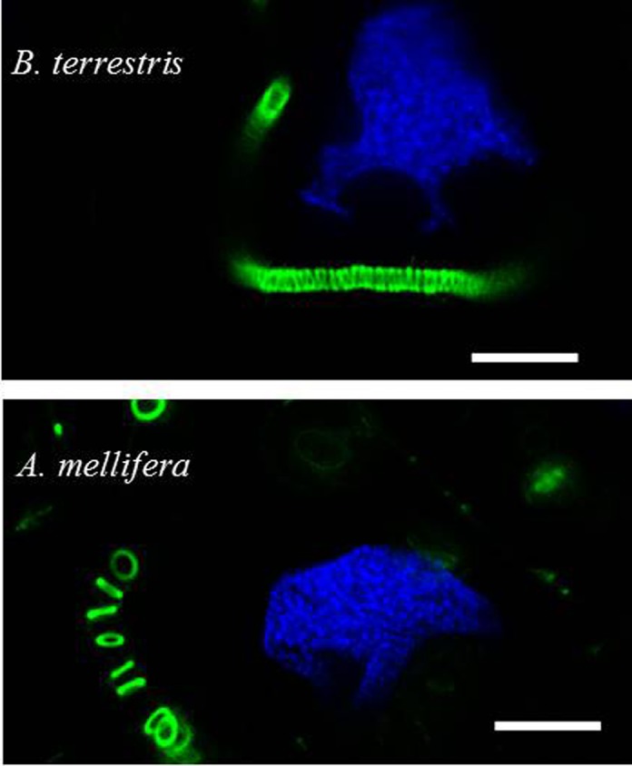
Dissected hypopharyngeal glands were treated with Hoechst stain (blue) and fluorescently labeled phalloidin (green). Actin rings of the bumblebee's end apparatus are smaller in diameter (∼2.0 µm), densely stacked, and slightly leaned to each other; those of honeybees have a larger diameter (∼3.1 µm) and do not contact each other. Scale bars: 10 µm.