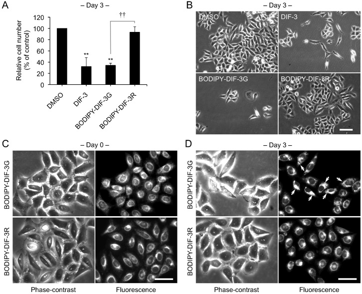
Fig. 2. Effects of BODIPY-DIF-3G and BODIPY-DIF-3R on HeLa cell growth, and cellular localization of the BODIPY-conjugated compounds.
(A) Cells were incubated for 3 days with 0.2% dimethyl sulfoxide (DMSO; vehicle) or 20 µM of BODIPY-DIF-3G or BODIPY-DIF-3R, and relative cell number was assessed. Mean values and s.d. (bars) of three independent experiments are presented. **P<0.01 versus DMSO control. ††P<0.01. (B) HeLa cells were incubated for 3 days with 0.2% DMSO or 20 µM of DIF-3, BODIPY-DIF-3G, or BODIPY-DIF-3R, and observed by using phase-contrast microscopy. (C,D) Cells were incubated for 0.5 h (C) or 3 days (D) with BODIPY-DIF-3G (20 µM) or BODIPY-DIF-3R (20 µM), washed free of the additives, and observed by using phase-contrast and fluorescence microscopy. Mitochondrial swelling (arrows) was induced in most of the cells treated with BODIPY-DIF-3G. Scale bars: 100 μm (B), 50 μm (C,D).

