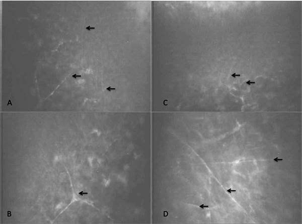
Figure 3.

Nerve regeneration into the central cornea of a typical cornea, as observed by in vivo confocal microscopy. (A, B) Unoperated control cornea showing the subepithelial zone and anterior stroma, respectively, at 9 months postoperative. (C, D) Corresponding zones from the contralateral implanted cornea. Nerves are indicated by the arrows.
