
Figure 1.
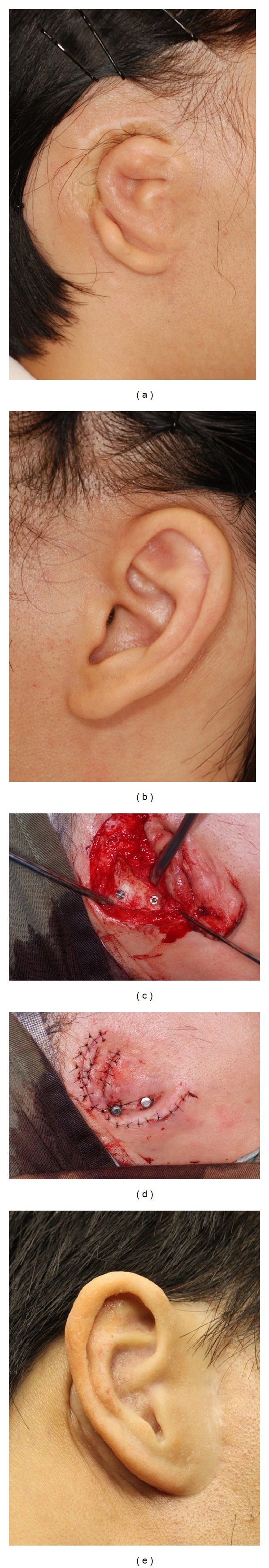
Case illustration of a patient with microtia. (a) Patient's right ear showing microtia. (b) Patient's left ear showing normal shaped ear. (c) Two implants were placed into the mastoid bone. (d) Wound closure with healing abutment exposed. (e) Patient's right ear showing the auricular prosthesis.