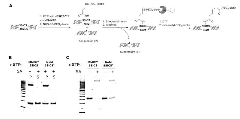
Figure 4.
(A) Immobilization of biotinylated dsDNA on streptavidin affinity resin. (B) Gel mobility assay of PCR amplicons labeled with NHS-SS-PEG4-biotin (P) compared to the unbound fraction remaining in the supernatant (S) after binding to the streptavidin solid support. Biotinylation levels are 53% for dMMO2A-d5SICS and 70% for dNaM-d5SICSA. A 100 bp DNA ladder is loaded in the leftmost lane. (C) Conjugation of dsDNA to iodoacetyl-PEG2-biotin after release from the streptavidin affinity resin via DTT treatment. Biotin incorporation levels are 89% for dMMO2A-d5SICS and 53% for dNaM-d5SICSA. A 50 bp DNA ladder is loaded in the leftmost lane. In (B) and (C), the faster migrating, strong band corresponds to dsDNA, while the slower migrating band corresponds to the 1:1 complex between dsDNA and streptavidin.

