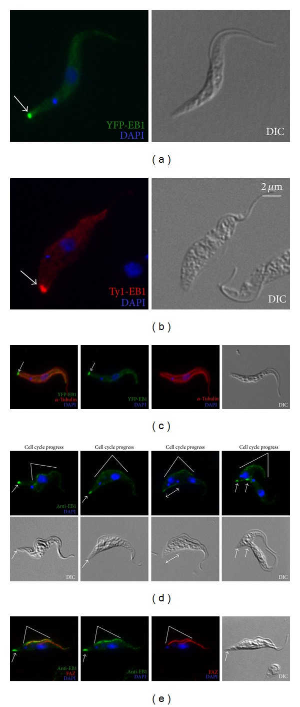
Figure 5.

Subpellicular microtubule plus end dynamics revealed by EB1. Cells stably expressing YFP-EB1 (a) or Ty1-EB1 (b) were fixed with cold methanol and labeled with DAPI for DNA. YFP-EB1 cells were also immunolabeled with anti-α-tubulin which revealed the total microtubule profile in a parasite cell (c). A polyclonal anti-EB1 was used to label microtubule plus ends throughout the cell cycle (d). Cells double labeled for anti-EB1 and FAZ revealed a possible nonspecific labelling of anti-EB1 along the FAZ region (e). Arrows, EB1 staining at the posterior tip of the cell; double headed arrow: elongated EB1 pattern during mitosis; white lines: possible nonspecific EB1 labelling near FAZ.
