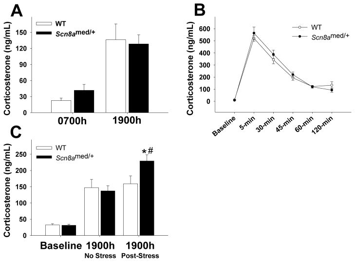
Figure 4. HPA axis function.
(A) Snc8amed/+ mice have normal diurnal fluctuations in plasma corticosterone levels, with similar corticosterone levels as WT mice during both the nadir (0700h) and peak (1900h) of the circadian HPA axis rhythm (time main effect: F(1,16)=34.011, p<0.001). (B) Plasma corticosterone levels before (baseline) and at 5, 30, 45, 60, and 120 min following a 20-minute restraint stress. There were no differences in the HPA axis response between Scn8amed/+ mice and WT mice (time main effect: F(5,130)=100.337, p<0.001). (C) Although there are no basal differences in plasma corticosterone levels prior to the stressor (baseline) between genotypes (t(20)=0.216, p=0.831), morning exposure to acute restraint stress increases corticosterone levels at 1900h in Scn8amed/+ mice, but not in WT littermates (stress main effect: F(1,20)=14.505, p<0.01; stress x genotype interaction effect: F(1,20)=8.605, p<0.01). *p<0.05 vs. WT within Post-Stress condition and #p<0.05 vs. No Stress condition within genotype, Tukey post hoc test. Error bars represent SEM. n=9–12 per group.

