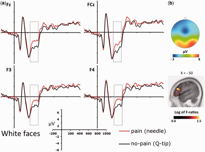
Fig. 3.
(A) ERPs recorded at a selection of frontal electrode sites (i.e. Fz, FCz, F3 and F4) relative to the two stimulation conditions (painful vs. nonpainful) for white/own-race faces. (B) Voltage topography of the N2–N3 activity recorded in the painful condition (upper panels) and source estimation of the N2–N3 activity in the painful vs. nonpainful conditions for white/own-race faces (lower panels).

