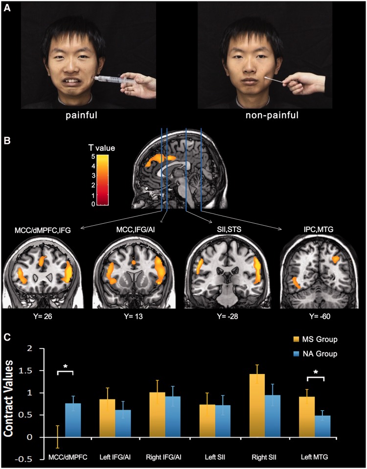
Fig. 1.
Illustration of stimuli and neural activity to perceived pain in others. (A) Illustration of the stimuli used in our study. Video clips showed painful faces receiving needle penetration or neutral faces with Q-tip touch. (B) Increased neural responses to painful vs non-painful stimuli during the pre-priming sessions. These were identified in the whole brain analysis in the MCC/dMPFC, AI/IFG, SII, STS, MTG and inferior parietal cortex (IPC). (C) Contrast values to painful vs non-painful stimuli during the post-priming sessions in MS and NA groups. NA group showed significantly greater MCC/dMPFC activity compared with MS group, whereas activation in the left MTG showed a reverse pattern.

