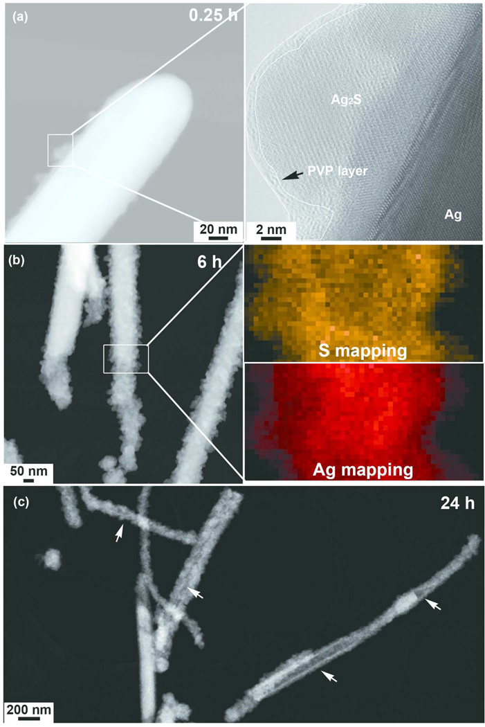
Figure 6.
HAADF-STEM images showing the morphological evolution and sulfidation process of AgNWs incubated in DCCM-1 medium for (a-left) 0.25 h, (b-left) 6 h and (c) 24 h. An HRTEM image (a-right) taken from the boxed area in Fig.6a-left shows the presence of a PVP layer(s) on sulfidized AgNWs surface. The PVP layer which shows a weaker contrast intensity and amorphous morphology is delineated using a white contour. The boxed area in Fig. 6b was further characterized by STEM-EDX Ag and S elemental mapping (Fig. 6b-right). The morphological evolution of the AgNWs in DCCM-1, as a function of time was characterized by HAADF-STEM. After incubation in DCCM-1 at 37 °C for 0.25 h, a thin surface layer of crystals was present (Fig. 6a-left). A HRTEM image of 24 h sample is presented in SI, Fig 13S.

