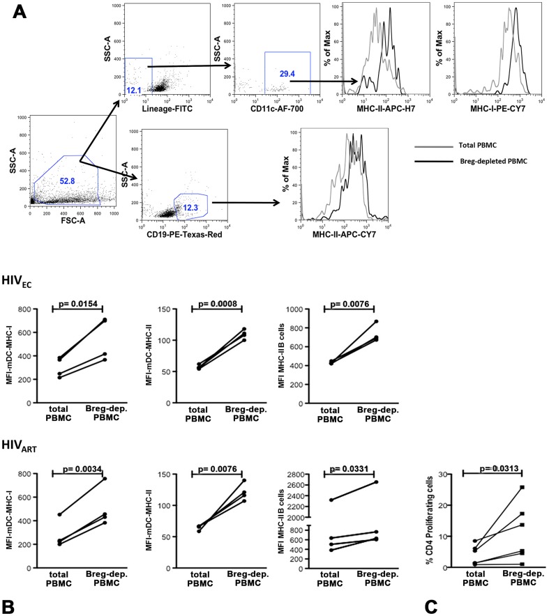
Figure 4. SAHA-treated Breg-depleted PBMC from HIVEC and HIVART exhibit upregulated expression of antigen-presenting molecules and proliferation of CD4+ T cells.
After 4 days in culture, (a) the expression of MHC-II and MHC-I/II on B cells and dendritic cells (LIN−CD11c+HLA-DR+) respectively was determined by flow cytometry in (b) SAHA-treated total or Breg-depleted PBMC from HIVEC, (n = 4, upper panel) and HIVART, (n = 4, lower panel). The gating strategy and representative histogram overlays are depicted in Figure 3a. (c) VPD450-proliferation dye labeled total or Breg-depleted PBMC were stimulated with SAHA (500 nM, Figure 1b right panel, n = 5) and after 4 days in culture proliferation of CD4+ T cells was determined by flow cytometry. p values for differences in CD4+ T cell proliferation as calculated by paired two-tailed t test (Graphpad Prism software) are indicated.

