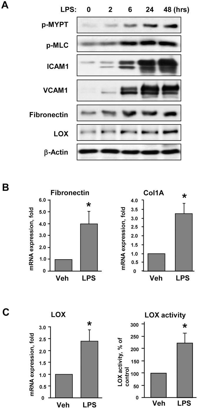
Figure 1. Characterization of LPS-induced activation of pulmonary EC.
Human pulmonary EC were grown on 2.8(200 ng/ml). A - Time-dependent phosphorylation of MYPT and MLC and expression of ICAM-1, VCAM-1, fibronectin and LOX was determined by western blot analysis. Equal protein loading was confirmed by membrane re-probing with β-actin antibody. B – Analysis of fibronectin and collagen 1A (Col1A) mRNA levels after 12-hr LPS treatment was performed by RT-PCR; C – Analysis of LOX mRNA levels and LOX activity in conditioned medium from control and LPS-stimulated (12 hrs) cells; *p<0.05.

