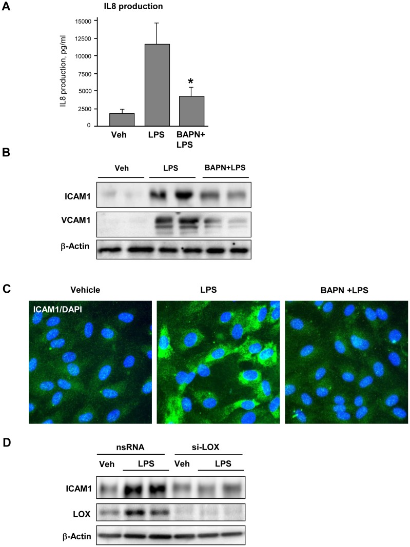
Figure 2. Effect of LOX inhibition on LPS-induced EC inflammatory activation.
Human pulmonary EC grown on 2.8(300 µM), and then stimulated with LPS (200 ng/ml) for 48 hrs with or without BAPN. A – IL-8 production by EC stimulated with or without BAPN was evaluated in conditioned medium by ELISA assay; *P<0.05. B – Expression of ICAM-1 and VCAM-1 was determined by western blot analysis with specific antibodies. C – ICAM-1 expression was examined by immunofluorescence staining of stimulated EC using ICAM-1 antibody (green). Counterstaining with DAPI (blue) was used to visualize cell nuclei. D – HPAEC were transfected with non-specific (nsRNA) or LOX-specific siRNA (si-LOX). ICAM-1 expression was determined by western blot. Equal protein loading was confirmed by membrane re-probing with β-actin antibody.

