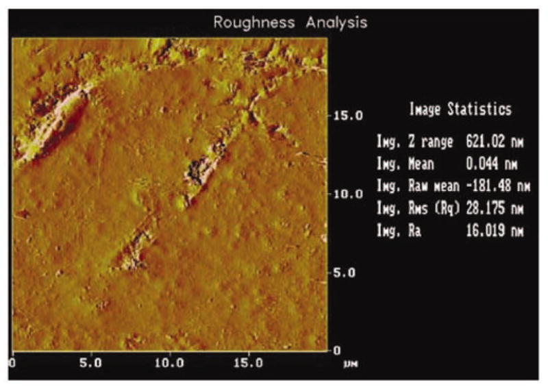
Figure 3.

Atomic force microscope imaging demonstrating the molding ability of a nine-micron-diameter quartz filament by optical laser two-dimensional representation with true statistical information acquired through the three-dimensional counterpart (x-y scales in microns). Rare surface imaging exposes a quartz filament damaged during mixing, placement, and mold separation. Fibrils extending upward provide most of the relief, appearing as white higher-modulus material outlined sporadically by black surrounding deeper defects compared with the large mass of dark greyish particulate-filled paste. Although damage creates an overall vertical Z-range distance of 621 nanometers, due to the ability of the pure quartz fiber to deform by compressing parallel against a surface and particulate paste to fill in space, the average surface roughness is still distinctively only 16 nanometers.
